
 打印
打印


Uranium is a naturally existing heavy metal with an abundance of about 2.3 μg/g in the Earth’s crust aggravated with the development of nuclear power and other anthropogenic activities. Its chemical toxicity of uranium mainly caused by uranyl ions (UO22+) poses health risks that can damage the kidney, bone, liver, brain, and lungs. To understand the cellular toxicity of uranium is necessary to better prevent and mitigate its toxic effects.
Due to the scarcity of investigation on non-uranium-accumulating mammalian organ cells, CIRP selected CHO-k1, a commonly used hamster ovarian epithelial cell line from ovary, to evaluate the behavior of uranyl ions in the cells and the impact on cell viability and morphology. The latter was analyzed by using scanning electron microscopy (SEM) and transmission electron microscopy (TEM). It also investigated the uptake and efflux process of uranium by CHO-K1 cells using inductively coupled plasma mass spectrometry (ICP-MS).
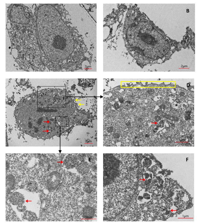
TEM analysis of CHO-k1 cells after 24 h of uranium exposure. Shown are representative images of (A) control (no uranium exposure), × 1.5 k; (B) 50 μ M
uranium exposure, × 2 k; (C) 300 μ M uranium exposure, × 1.5 k; (D) uranium precipitates (intracellular), × 5 k; (E) uranium precipitates (intracellular), × 12 k; and (F) uranium precipitates (extracellular), × 6 k. Red arrow indicates uranium precipitates; yellow arrow indicates leaked intracellular substances; yellow box indicates rough cell membrane.
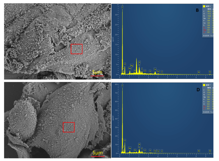
SEM analysis of CHO-k1 cells after 24-h uranium exposure. (A) SEM image of cells exposed to 50 μ M uranium, × 3 k, and (B) EDX spot analysis of the selected area, as indicated by the box. (C) SEM image of cells exposed to 300 μ M uranium, × 3 k, and (D) EDX spot analysis of the selected area, as indicated by the box.
The cytotoxicity of uranium in CHO-k1 cells was found to be dose related and was abated by lowering the temperature or by adding an endocytosis inhibitor. The cells were blocked at G1 phase and the percentage of adherent cells was significantly decreased, indicating significantly inhibited cell proliferation. IPC-MS analysis revealed for the first time that the efflux of uranium was a very rapid process. In addition, it was observed that uranium formed needle-like precipitates not only in the extracellular regions but also in lysosomes and intracellular vesicles, which suggests that mineralization may be one of the fates of accumulated uranyl ions, especially at high concentrations. Finally, gene transcriptome analysis indicated that uranium exposure induces the transcription changes of metabolism and redox related genes. These results collectively provide insights into the mechanism of uranium cytotoxicity at the cellular and molecular levels and suggest that more careful and elaborate studies should be conducted on the interaction of uranium and cells.
Contact: official@cirp.org.cn